This chapter will discuss Anatomical Planes, Extremities and Subdivisions, Positional Terms, Respiratory System, Circulatory System, Digestive System, Urinary System, Female Reproductive System, Male Reproductive System, Nervous System, Endocrine System, Musculoskeletal System, Major areas of the skeleton, Major Types Of Muscle and The Skin with its physical features all in complete detail.
Anatomical References / Human Body Systems & Organs
OBJECTIVES:
- Upon completion of this lesson, you will be able to:
- Define anatomical position.
- Identify and describe the three anatomical planes.
- Identify the five regions of the human body.
- List the five body cavities and the organs they contain.
- Describe the location of a wound on a patient using anatomical references
- List the body system and identify the main organ included in this system.
- Name the four Abdominal quadrants.
- Identify the main Internal organs located in each Abdominal quadrant.
1. Anatomical Position
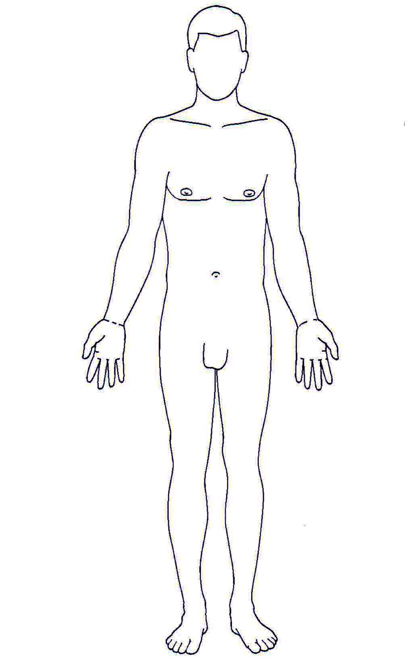 Definition: Standing erect with arms down at the sides, palms facing forward. “Right” and “left” refer to the patient’s right and Left.
2. Conventional References
2.1 Anatomical Planes
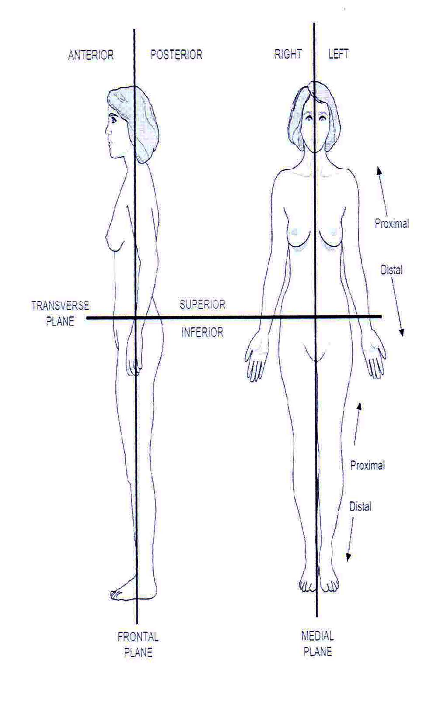
The anatomical planes refer to imaginary planes that divide the body in two halves, in different orientations. Write in the descriptions of the three anatomical planes below:
- Medial plane:
The median air plane also called a mid-Sagittal atmosphere plane could be utilized to describe the Sagittal air plane as it bisects your human anatomy upright by utilizing the midline labeled by the navel, dividing the body completely in left and side that’s right.
- Transverse plane:
The transverse plane (also referred to as the horizontal plane, axial plane, or trans-axial plane) is an imaginary plane that divides the body into superior and inferior parts.
- Frontal plane:
The coronal plane ( frontal plane) see in (vertical) divides the human body in to dorsal and ventral (back and front, or posterior and anterior) segments.
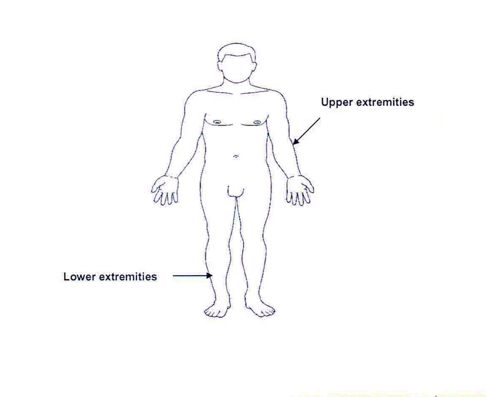
2.2 Extremities and Subdivisions
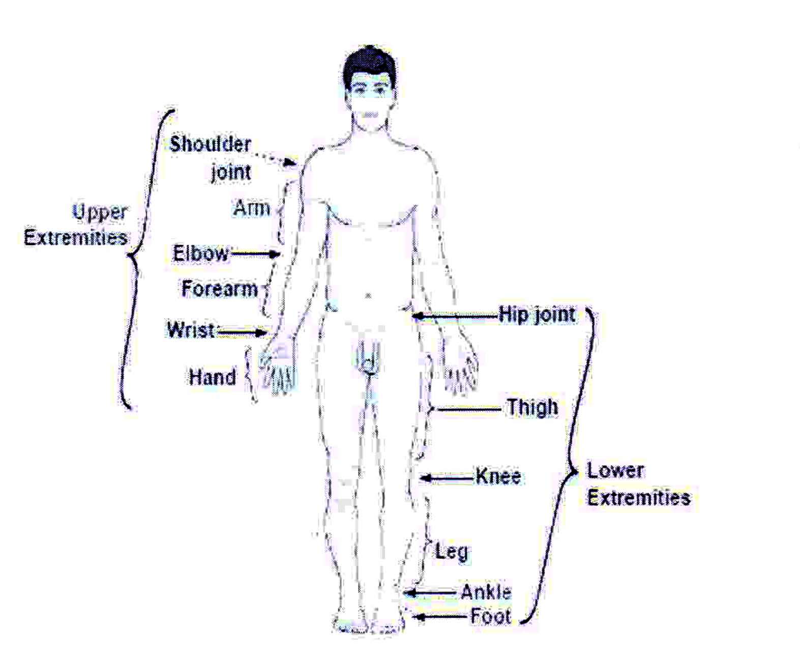
Proximal: Means close, or closer to the point of reference given. Distal: Means distant, or farther away from the point of reference given.
2.3 Positional Terms
Prone:
Supine:
Lateral recumbent or “recovery”:
Extremities and Subdivisions
3. Body Regions
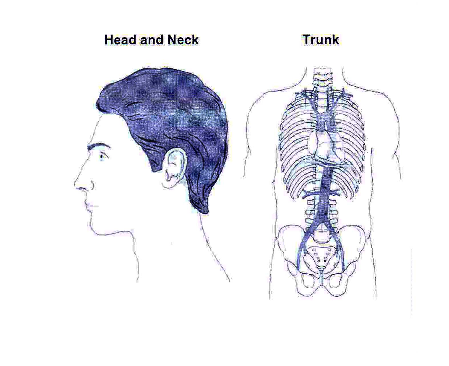
Body Regions
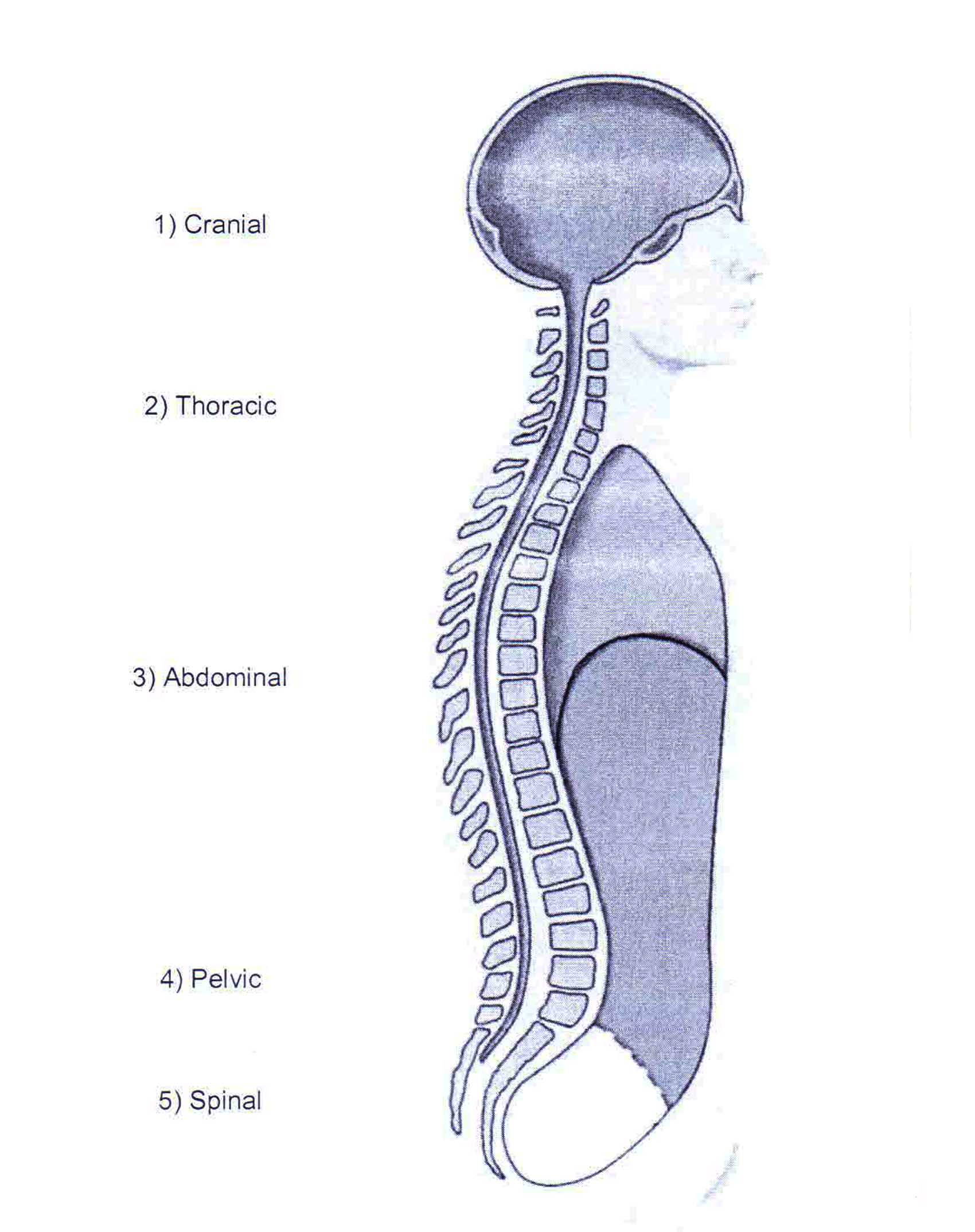
4. Body Cavities
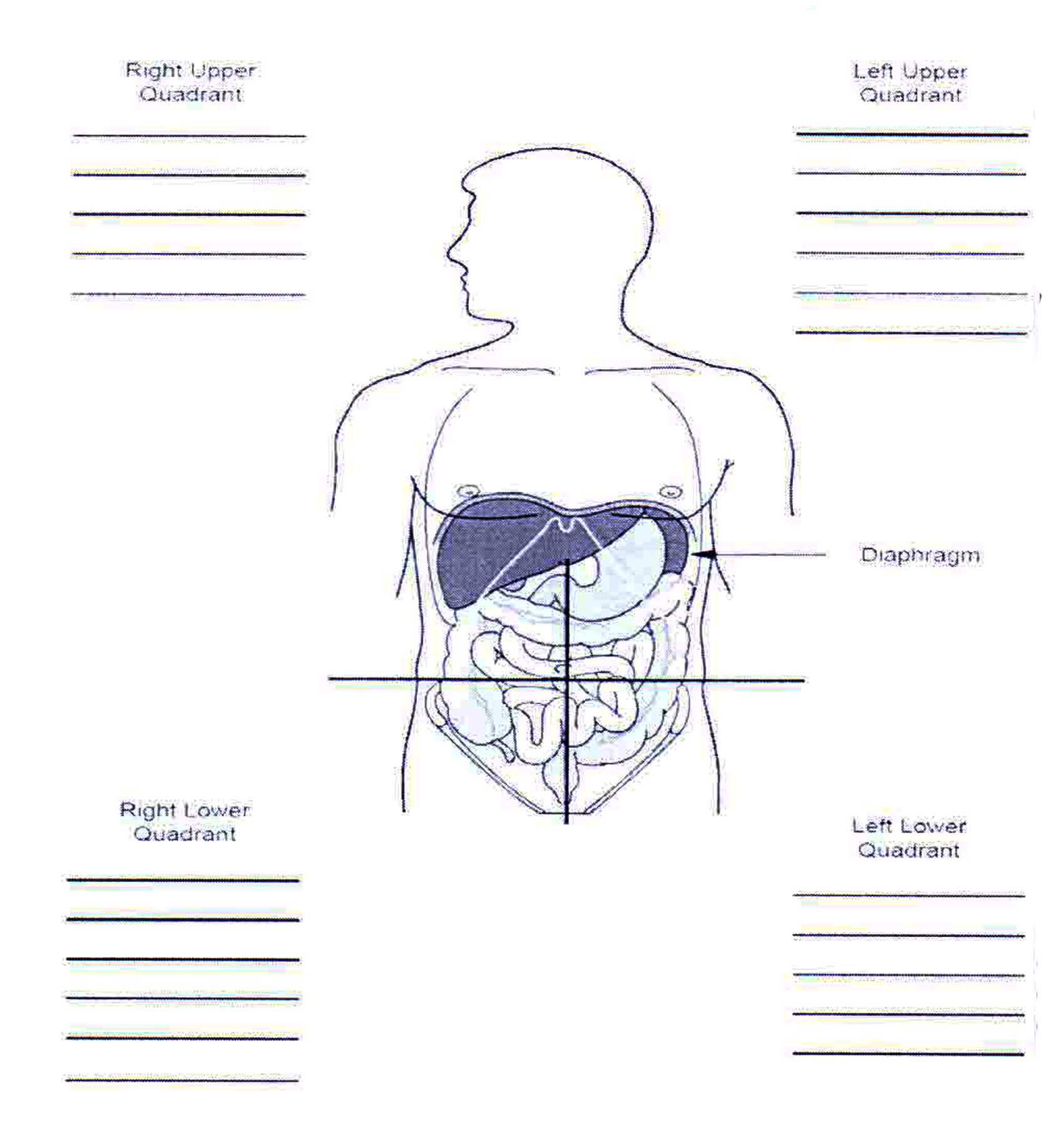
5. Abdominal Quadrants and Organs
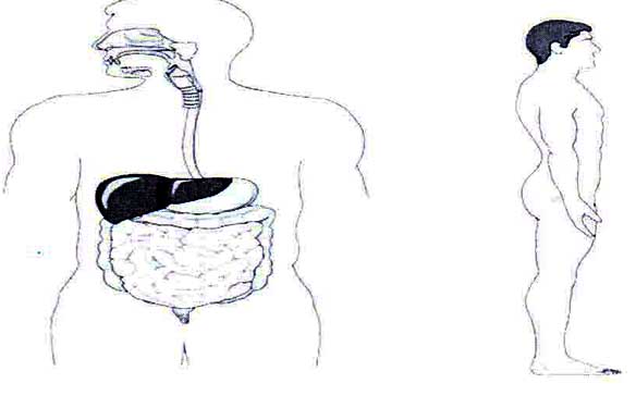
Organs in the midline area is lined with a membrane and include the stomach, lower part of the esophagus, small and large intestinal tracts (except sigmoid and rectum), spleen, liver, gallbladder, pancreas, adrenal glands, kidneys, and ureters.
Hollow abdominal organs:
The hollow abdominal organs have an esophagus, small intestine, colon (large intestine), and stomach.
Solid abdominal organs:
The solid organs include liver, adrenals, pancreas, spleen, kidneys, ovaries and uterus.
Location of kidneys:
The left kidney is situated slightly more superior than the right kidney because of to the larger size of the liver on the right side of the human body.
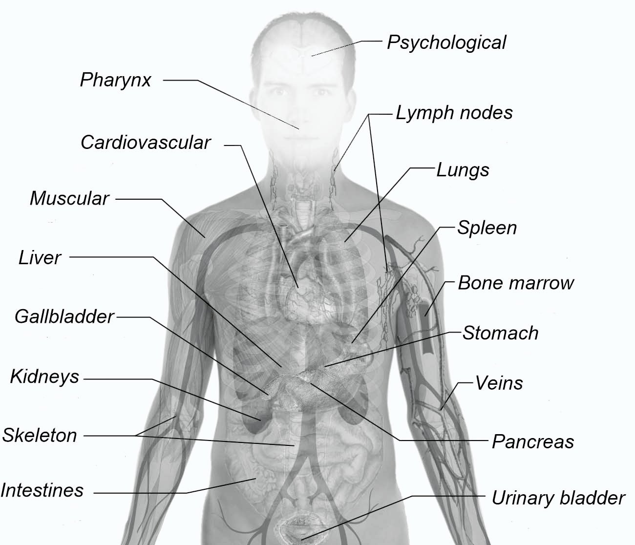
6. Body Systems
6.1 Respiratory System
The function of the respiratory system is to deliver oxygen to the body and to remove carbon dioxide from the body. The air passing into and out of the lungs is known as respiration. Breathing in is called inspiration or inhaling and breathing out is called expiration or exhaling. While breathing or during the process of inspiration, the muscles of the thorax contract, moving the ribs outward and up. 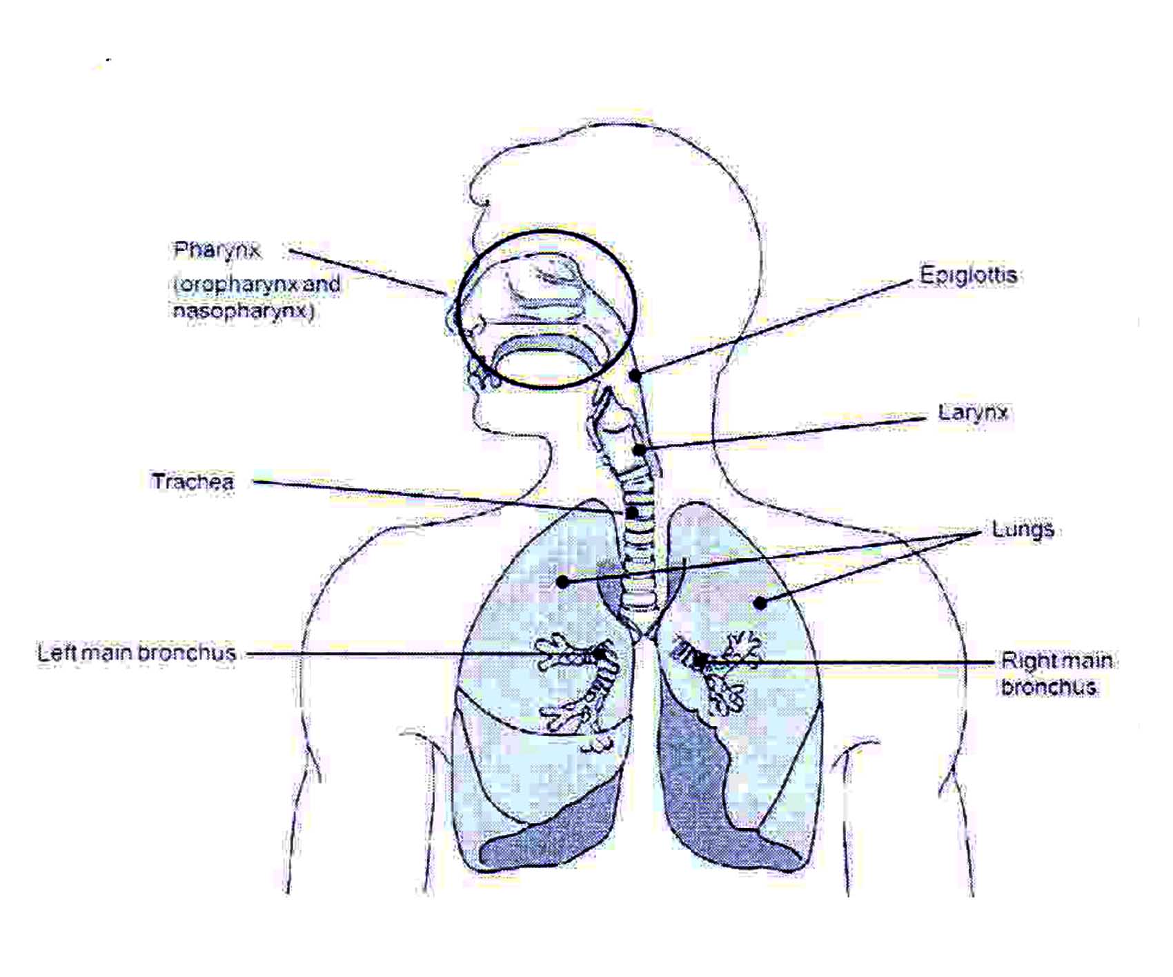 The diaphragm contracts and lowers. This process expands the chest cavity and causes air to flow into the lungs. During exhalation, the opposite occurs. The muscles of the chest relax and cause the ribs to move inward. At this time, the diaphragm relaxes and moves up.
The respiratory system is made up of the organs that allow us to breathe. Air enters in through the nose and the mouth. The area behind the mouth and nose is called the pharynx which is divided into the oropharynx and the nasopharynx (windpipe). The trachea is the air passageway to the lungs. The epiglottis is a leaf-shaped structure that keeps foreign objects from entering the trachea during the swallowing process. The trachea splits into two bronchi. These air passages become smaller and smaller until they reach the alveoli, where carbon dioxide and oxygen are exchanged with blood.
6.2 The Circulatory System
 The cardiovascular system consists of the and. The heart is a muscular organ, approximately the size of a fist, and is located in the thoracic cavity behind the sternum and between the lungs. The coronary arteries are special arteries that supply blood to the heart muscles themselves. The function of the respiratory system is to deliver oxygen to the body and to remove carbon dioxide from the body. Air passing into and out of the lungs is known as respiration.
Breathing in is called inspiration or inhaling and breathing out is called expiration or exhaling. While breathing or or during the process of inspiration, the muscles of the thorax contract, moving the ribs outward and up. The diaphragm contracts and lowers. This process expands the chest cavity and causes air to flow into the lungs. During exhalation, the opposite occurs. The muscles of the chest relax and cause the ribs to move inward.
See also: EMS & MFR
At this time, the diaphragm relaxes and moves up. The function of the heart is to. Theheart receives oxygenated blood from the lungs and pumps it to the body through the arteries. The heart receives, from the veins, the blood that has circulated through the body and pumps it to the lungs to be oxygenated once again.
A system of one-way valves keeps the blood flowing in the right direction and prevents it from flowing backward.
6.3 Digestive System
 The system that is digestive of the alimentary tract (meals passageway) and extra organs. The primary function of the digestive system is to consume food and get rid of waste. Digestion is composed of two processes: mechanical and chemical. The mechanical process includes chewing, swallowing, the rhythmic movement of matter through the tract, and defecation (the
elimination of waste). The chemical process consists of breaking down food into simple components that can be absorbed and used by the body.
Excluding the mouth and the esophagus, the organs of the digestive system are in the abdomen. These organs include the stomach, pancreas, liver, gallbladder, small intestine, and large intestine.
6.4 Urinary System
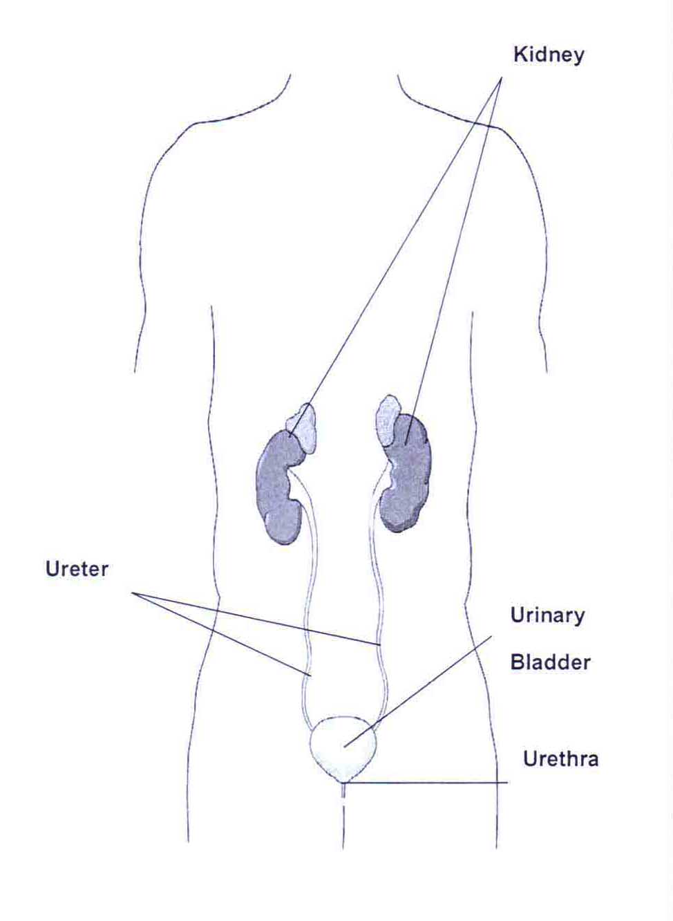
The system is urinary and excretes waste from the body. It is comprised of two kidneys and two ureters, one urinary bladder and one urethra. The ureters simply take urine through the kidneys to the part that is next of system-the the bladder. The bladder stores urine until it passes away through the urethra and is excreted through your body.
6.5 Female Reproductive System
 The reproductive system of the female consists of two ovaries, two fallopian tubes, the uterus, the vagina and external genitals.
The female reproductive system provides the egg (ovum) which is fertilized by the male’s sperm.
6.6 Male Reproductive System
 The reproductive system of the male consists of two testes, the seminal duct, accessory glands, and the penis. The male reproductive system provides the sperm which fertilizes the female’s ovum.
6.7 Nervous System
 The nervous system is composed of the brain, the spinal cord, and nerves. The nervous system has two major functions: communication and control. This system lets a person be aware of and react to the environment. It coordinates the body’s responses to stimuli and keeps body systems working together.
The stressed system has three main components: the central stressed system, the peripheral nervous system plus the autonomic nervous system. The nervous system comprises the brain therefore the spinal-cord. The peripheral system is nervous of the nerves. The autonomic system which is nervous functions throughout the human anatomy.
6.8 Endocrine System
 The endocrine glands regulate the body by secreting hormones directly into the bloodstream. These glands affect physical strength, mental ability, stature, reproduction, hair growth, voice pitch, and behavior. The secretions from these tiny glands can affect how people think, act and feel. Each gland produces one or more hormones. Some of the glands in the endocrine system are the thyroid, parathyroids, adrenals, ovaries, testes, and the pituitary.
6.9 Musculoskeletal System
The musculoskeletal system is made up of the skeleton and muscles. This system helps to give the body shape and to protect internal organs. Muscles also provide for movement.
The skeleton shapes the human body with its bony framework. The bone consists of living cells and nonliving matter. The nonliving matter contains calcium compounds that help make the bone hard and rigid. Without bones, the body would collapse. The skeleton is
held together mainly by ligaments, tendons and layers of muscle.
The three kinds of joints are immovable like the skull, slightly movable like the spine, and freely movable like the elbow or the knee.
Major areas of the skeleton
The skull has several broad, flat bones that form a hollow shell. The top, including the forehead, back, and sides of this shell make up the cranium.
The spine houses and protects the spinal cord. The spinal column is the main supportive structure that is bony of the body and consist of 33 bones called vertebrae. The spine is divided into five major sections: the cervical spine, the thoracic spine, the lumbar spine, the sacrum, and the coccyx.
The thorax, or rib cage, protects the heart and lungs, vital organs of the body. They are enclosed by 12 pairs of ribs and are attached at the back to the spine. The top 10 pairs are also attached in the front to the sternum or breastbone. The lowest portion of the sternum is called the xiphoid process.
The pelvis, or hip bones, consists of the ilium, pubis, and ischium. Iliac crests fare rom the “wings” of the pelvis. The pubis is the anterior portion of the pelvis. The ischium is the posterior portion. The shoulder girdle consists of the clavicle (collarbone) and the scapulae (shoulder blades).
The upper extremities extend from the shoulders to the fingertips. The arm (shoulder to elbow) has one bone known as the humerus. The bones in the forearm (elbow to wrist) are the radius and the ulna.
The lower extremities extend from the hips to the toes. The bone in the thigh, or upper leg, is known as the femur. The bones in the lower leg (knee to ankle) are the tibia and fibula. The kneecap is called the patella.
Major Areas of the Skeleton
Major Types Of Muscle
 Skeletal muscle, or voluntary muscle, makes possible all deliberate acts like walking and chewing.
Smooth muscle, or involuntary muscle, is made of longer fibers and is located in the walls of tubelike organs, ducts, and blood vessels and forms much of the intestinal wall. A person has little or no control over this type of muscle.
Cardiac muscle makes up the walls of the heart. This muscle can stimulate itself into contraction, even when disconnected from the
brain.
6.10 The Skin
The skin protects the body from the world that is outside. It also protects the deep cells from injury, drying out, and invasion by germs and other bodies that are foreign. The skin also helps to regulate the human anatomy temperature, aids in eliminating water and salts that are various, and helps to avoid dehydration. The skin also acts as the receptor organ for touch, discomfort, heat, and cool.

The epidermis is the outermost layer of the skin and contains cells that give it color. The dermis, or second layer, contains a vast network of blood vessels. The deepest layers of the skin contain hair follicles, sweat and oil glands, and sensory nerves. Just under the skin is a layer of subcutaneous fatty tissue.
Aroused Questions from this Chapter:
1. Define anatomical position
Ans: The position of an animal’s body at that the totality of its muscle groups have reached their stress that is cheapest. The place that is an anatomical person standing erect, with feet dealing with ahead, the arms at the sides, palms of the hands facing the interior, and hands pointing along for individual beings.
2. Describe the location of a wound on a patient using anatomical references. Identify the approximate location of the injuries indicated by the circles. (Respond on the following page).
Ans:
3. List the Four regions of the human body on a skeletal model.
he skeletal system is composed of four primary fibrous and mineralized tissues that are conjunctive bones, ligaments, tendons, and joints. Bone: A rigid form of conjunctive tissue that is part of the skeletal system of vertebrates and it is composed mainly of calcium mineral.
4. List five cavities of the body and the organs they contain.
- Cranial cavity. Housing the human brain and the pituitary glad.
- Spinal cavity. Housing the spinal cord.
- Thoracic cavity. Housing the lungs.
- Abdominal cavity. Housing the major digestive organs.
- Pelvic cavity. Lodging the urinary and reproductive organs.

Alex Smith, a seasoned medical technician with 15 years in ambulance services, writes crucial first-aid tips and emergency care insights on arescuer.com.

